An Unusual Case of Syncope
Ashley D Nelson, MD
Pulmonary and Critical Care Fellow
University of Utah School of Medicine
History of Present Illness: A 56 year old female presented to the hospital for an episode of syncope. Her syncope occurred earlier in the day while she was going to the bathroom. She felt dizzy, and then briefly fainted. Her mother, who is also her caregiver, witnessed the episode. It lasted for a few seconds and the patient quickly returned to her baseline level of consciousness. She has had similar episodes in the past.
Past Medical History: Mitral valve endocarditis complicated by an embolic cerebellar stroke one year ago. She continues to have nausea, vertigo, poor oral intake with subsequent weight loss and failure to thrive. She also has chronic kidney disease and coronary artery disease.
Physical Exam:
Upon presentation her vital signs were normal. She had a normal physical exam.
EKG: Qtc 480 msec, otherwise normal.
Initial labs: WBC- 3 x 10³/µL, Hemoglobin- 10 g/dl, Troponin I- 0.37 ng/mL, Creatinine- 2.2 mg/dl (baseline is 2), the rest were within normal limits.
Imaging: A CT head and CXR were normal.
Given her history of endocarditis, an echocardiogram was performed.
Question: What is the most likely diagnosis?
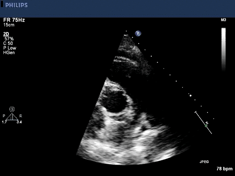
Pulmonary Saddle Embolus
The echocardiogram shows an image of the right ventricular outflow tract (RVOT) and the bifurcation of the main pulmonary artery. The aorta is seen on the left side of the image. At the bifurcation of the pulmonary artery, a mobile hyperechoic mass consistent with pulmonary embolus is seen. Interestingly, a VQ scan performed the subsequent day demonstrated a very low probability of pulmonary embolism.
This case illustrates a few pitfalls in an unusual presentation of a PE. First, the patient’s pretest probability was low (Wells score of 1.5 for immobility), and even if the D dimer were elevated, the very low probability VQ scan would have argued for no further treatment. However, it is important to recognize that even with a very low probability VQ scan, there can be up to a 10% probability of a PE (1). Second, echocardiography is not a standard method to diagnose a PE. However, echo can often provide useful supplementary information regarding right heart hemodynamic changes that can occur with a PE, such as right ventricular dysfunction, or elevated right ventricular systolic pressure. Additionally, echocardiography is highly specific if a thrombus is visualized in the heart or pulmonary artery (>90%). However, the corresponding sensitivity is low (2).
The patient was treated with anticoagulation and her syncope episodes resolved.
References:
- Value of the ventilation/perfusion scan in acute pulmonary embolism: Results of the prospective investigation of pulmonary embolism diagnosis (PIOPED). JAMA : The journal of the American Medical Association 1990; 263: 2753-2759.
- Kearon C. Diagnosis of pulmonary embolism. CMAJ : Canadian Medical Association Journal. 2003; 168: 183-194.
05/2015


