Trouble on the Horizon
Andrew Badke, MD
Pulmonary and Critical Care Medicine Fellow
University of Utah School of Medicine
Case:
A 40 year old female has been experiencing progressive dyspnea and exertional pre-syncopal episodes over the course of several weeks. Her history is significant for inflammatory bowel disease and obesity.
Vitals: Blood pressure: 136/86, Heart rate: 106, Respiratory rate: 26, pulse oximetry 83% on room air
Physical exam is unremarkable. D-dimer and BNP are elevated. Troponin is normal. EKG has evidence of right heart strain.
A CT confirms a diagnosis of bilateral pulmonary emboli.
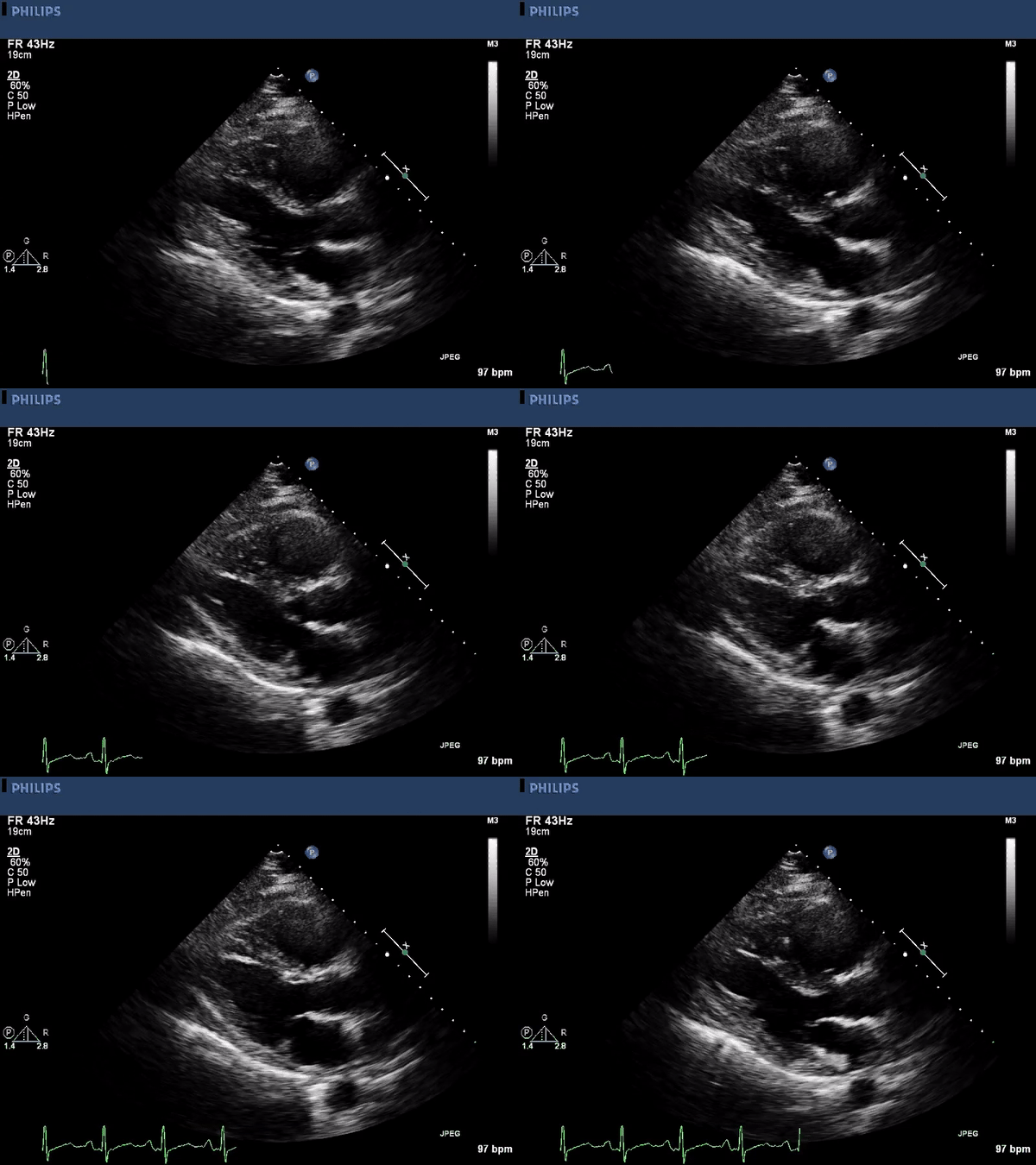
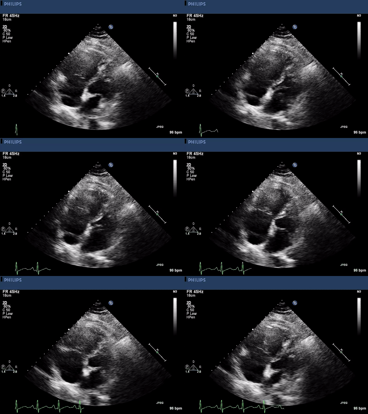
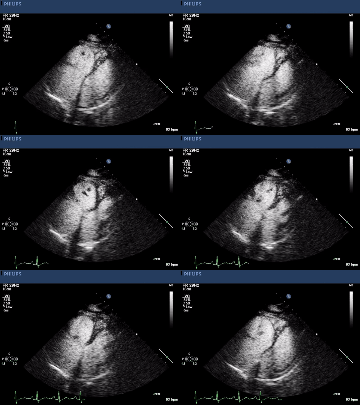
An echocardiogram is performed:


The patient is started on a Heparin infusion in the emergency department.
What is the next step in management?
- Discharge the patient with a prescription for low molecular weight heparin and close follow up
- Continue heparin infusion and admit the patient to the medical intensive care unit
- Emergently place an IVC filter to prevent further embolization
Answer B: Management should take place in the intensive care unit while continuing anticoagulation. Thrombolytic therapy or surgical embolectomy should also be considered.
The echocardiographic images above show a large right ventricular thrombus. The first image is a parasternal long axis view highlighting the right ventricular inflow tract. A mobile thrombus is seen near the anterior border. The other two images are using an apical approach showing the right and left ventricle. Again a mobile the thrombus is seen within the right ventricle. Contrast is used to enhance the thrombus borders.
Echocardiography can be a helpful tool when managing patients with pulmonary embolism. The presence or absence of right ventricular (RV) dysfunction can readily assist with triage and prognostication. Concerning findings for RV dysfunction on echo include: RV dilation, increase in RV-LV diameter ratio, hypokinesis of the RV free wall, increased tricuspid regurgitant jet velocity, or a decreased TAPSE. There tends to be wide variability in clinical outcomes when RV dysfunction is present on echo, which can make determining the clinical significance difficult. However, patients with none of the above findings are typically very low risk for developing complications and can safely be triaged to a lower level of care or even be managed in an outpatient setting.1
In addition to assessing right ventricular function, echocardiography can also identify a right-to-left shunt through a patent foramen ovale, or the presence of a RV thrombus. In fact, the appearance of a thrombus in the right sided chambers of the heart can be a relatively common finding. The literature suggests that roughly 4-18% of patients will have a thrombus on TTE after a pulmonary embolism has been diagnosed.2,3 These patients have a significantly higher mortality rate than those with pulmonary embolism alone. In a 2002 meta-analysis, the mortality rate exceeded 25%, and without treatment was 100%.3
There are two patterns of right sided thrombi that have been described4. Type A is a highly mobile serpiginous, or worm-like thrombus. It is hypothesized that these clots have developed in the lower extremities and have embolized to the right chambers of the heart; otherwise known as a “thrombus in transit”. They can often be seen traversing through valves or even through a patent foramen ovale. Type A thrombus have been associated with a poor prognosis, and over 40% of patients with this type will have a thrombus-related death.4 Type B thrombi are more broad based and typically develop in the right heart itself. They are most often seen in patients with low cardiac output or dilated cardiac chambers.5 Patients with type B thrombi have a better prognosis and mortality rate in this group has been reported around 4%.4 There is a small subset of patients that fall between these two types of thrombi. The thrombus appears highly mobile, but not serpiginous. When analyzed, these in-between thombi were found to have intermediate mortality risk compared to the other two types.4
Treatment for right heart thrombi remains controversial. Because of the high mortality that has been associated with these cases, it is often felt that anticoagulation alone may be inadequate. In 1989, a meta-analysis found no significant difference between anticoagulation, thrombolytic therapy, or surgery in regards to mortality.4 However, in 2002 a separate meta-analysis found a significant advantage for thrombolytic therapy over anticoagulation alone or when combined with surgery. The study reported an odds ratio for mortality of 0.33 with thrombolytic therapy compared to anticoagulation, and 0.86 compared to surgery.2 Since there are no large randomized studies evaluating optimal treatment, clinical judgment must be used to determine the optimal therapy.
This patient had an intermediate-type thrombus with elements of type A and B. Given her clinical stability, she was initially monitored on heparin therapy alone for two days. She had a repeat echocardiogram and transesophageal echocardiogram which then demonstrated increasing size of the thrombus. She was ultimately taken to the operating room where she had an embolectomy of the right ventricle and left main pulmonary artery. She required a right ventricular assist device following the operation due to right ventricular failure after embolectomy.
References
- Members AF, Konstantinides SV, Torbicki A, Agnelli G, Danchin N, Fitzmaurice D, et al. 2014 ESC Guidelines on the diagnosis and management of acute pulmonary embolism. European Heart Journal. 2014 Nov 14;35(43):3033–73.
- Torbicki A, Galié N, Covezzoli A, Rossi E, De Rosa M, Goldhaber SZ. Right heart thrombi in pulmonary embolism: Results from the international cooperative pulmonary embolism registry. Journal of the American College of Cardiology. 2003 Jun 18;41(12):2245–51.
- Rose PS, Punjabi NM, Pearse DB. Treatment of right heart thromboemboli. Chest. 2002 Mar 1;121(3):806–14.
- Kronik G. The European Cooperative Study on the clinical significance of right heart thrombi. European Heart Journal. 1989 Dec 1;10(12):1046–59.
- Ferrari E, Benhamou M, Berthier F, Baudouy M. Mobile thrombi of the right heart in pulmonary embolism: Delayed disappearance after thrombolytic treatment. Chest. 2005 Mar 1;127(3):1051–


