The Breathing Pause
Biot’s Breathing Pallav Halani, MD 1 and Bradley Vaughn, MD 1
1Division of Sleep Medicine, Department of Neurology, University of North Carolina, Chapel Hill, North Carolina
Case
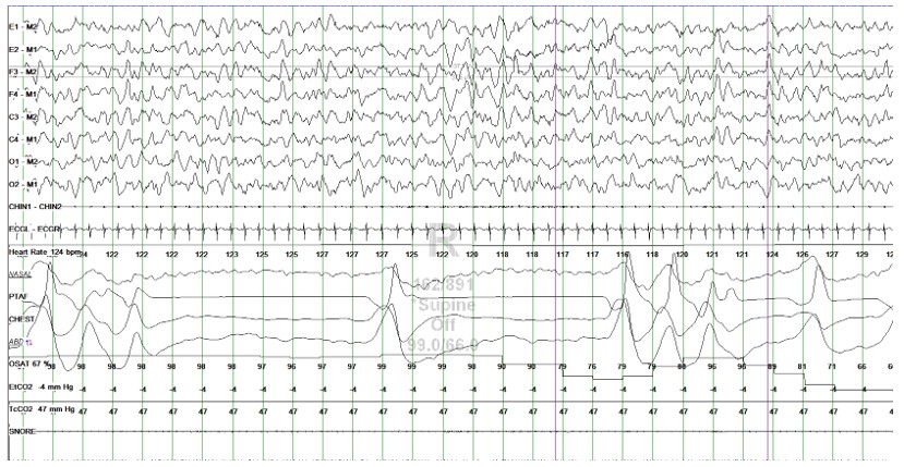
A nine month-old former 24 week preemie female twin with a history of bronchopulmonary dysplasia with hypoxemia on 0.5LPM oxygen supplementation, GERD, right grade II and left grade I perinatal intraventricular hemorrhage, neonatal hypertension, and stage II retinopathy of prematurity presents to our institution for an oxygen titration sleep study. Abnormal breathing pattern was seen during the polysomnography, and a representative 60-second epoch is pictured below.
Overall apnea hypopnea index (AHI) without supplemental oxygen was 54.9/h which reduced to 2.3/h with 0.5L supplemental oxygen. The respiratory channels showed rapid shallow breathing alternating with irregular periods of apnea lasting 7-8 seconds, and shorter pauses both associated with oxygen desaturations. As in the figure below the apnea was interrupted by single or multiple irregular breaths. This breathing pattern resolved with supplemental oxygen.
Question
What is your diagnosis?
- Central apnea
- Cheyne- Stokes respiration
- Biot’s breathing
- Cluster breathing
C. Biot’s breathing
Discussion
Camille Biot, a French physician first described Biot's breathing pattern in 1876. While studying the breathing pattern now known as Cheyne-Stokes respiration, Dr. Biot made observations in a 16-year-old child with tuberculous meningitis. He described this breathing pattern as lacking crescendo–decrescendo morphology, instead being completely irregular with varying periods of apnea. The breathing pattern shown in his original tracing demonstrated ataxia of respiration and not regular phasic breathing pattern.
"This irregularity of the respiratory movements is not periodic, sometimes slow, sometimes rapid, sometimes superficial, sometimes deep, but without any constant relation of succession between the two types, with pauses following irregular intervals, preceded and often followed by a sigh more or less prolonged." 2
Biot’s breathing pattern has been described to be associated with lesions of brainstem, namely upper medulla. Brain imaging should thus be considered in the patient’s presenting with Biot’s respiration.
The irregularity is a key differentiation between Biot’s breathing and the other answer choices. In cluster breathing, a series of breaths are separated by regular intervals of pauses, while in Cheyne Stokes breathing, a crescendo-decresendo pattern is seen. Central apnea lacks the pattern of irregularity noted in the patients with Biot’s breathing.
References
-
Powell, R Douglas. Biot on Cheyne Stokes The London Medical Record. 1877 Feb 15; 4:6566.
-
Biot Contribution a l’etude du phenomene respiratoire de CheyneStokes. Lyon Med. 1876;23:517528, 561-567.
-
Fisher CM. The neurological examination of the comatose patient. Acta Neurol Scand. 1969;45(S36):5-56.
-
Wijdicks EF. Biot's breathing. J Neurol Neurosurg Psychiatry. 2007;78(5):512-513.


