Stress while vacationing in the mountains
Thomas W. DeCato, MD
Pulmonary and Critical Care Medicine Fellow
University of Utah School of Medicine
Presentation:
A 53 year-old woman presented with worsening dyspnea over a 3-day period after traveling from sea level to an elevation of 7,000 feet. On the day of admission, she developed increased work of breathing and chest tightness. She denied fevers, chills or other new infectious symptoms. Her past medical history was significant for paroxysmal atrial fibrillation, mild intermittent asthma, and recent receipt of antibiotics for sinusitis. She was taking hormone replacement therapy for menopausal symptoms.
Physical Exam:
T 36°C, BP 100/50, HR 130, RR 32, SpO2 75% on ambient air
Heart rhythm was irregularly irregular. Mild accessory muscle use. Crackles were present at the bases bilaterally. Lower extremity edema was noted bilaterally.
Relevant Hospital data:
WBC 13 k/µL, Sodium 118 mg/dl, troponin 0.28 ng/mL, BNP 2565 pg/mL. CT angiogram showed bilateral pleural effusions and no pulmonary embolism.
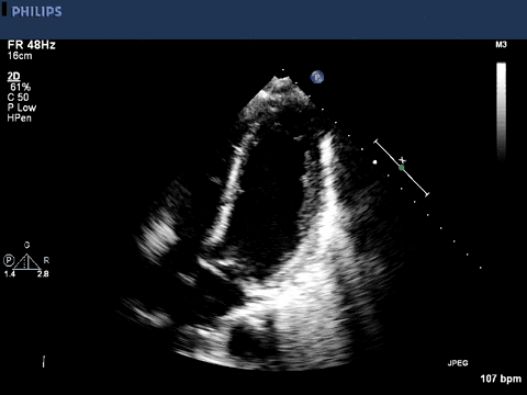
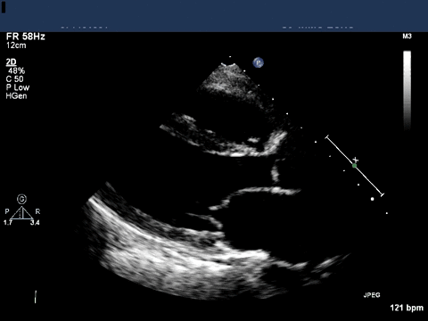
A transthoracic echocardiogram was obtained.
Question:
What is the most likely diagnosis?
Takotsubo Cardiomyopathy
Image 1 of the echocardiogram is an apical 2-chamber view showing apical ballooning of the left ventricle with hyperkinetic basal segments. Image 2 is a parasternal short axis view showing the same findings. Takotsubo cardiomyopathy, a specific presentation of stress cardiomyopathy, is an acute and reversible disorder of left ventricular (LV) dysfunction. Classically, echocardiography shows ballooning of the apex with apical akinesis and basal hyperkinesis (type I) of the LV. Midventricular ballooning, cardiomyopathy with apical hypercontractility, basal ballooning, localized type (types II-V) have also been recognized. The name takotsubo is taken from a pot that Japanese fisherman use as an octopus trap, which has a similar appearance.
Common presentation includes acute onset chest pain, ST segment changes on EKG concerning for acute coronary syndrome, and dyspnea. Arrhythmia (up to 8%) and congestive heart failure (up to 28%) may be evident on presentation as they were in our patient. Cardiogenic shock may be present in up to 5% of patients. Many diagnostic criteria have been proposed, and this remains a diagnosis of exclusion. Critical illness can also be associated with acute coronary syndrome, making it difficult to differentiate stress cardiomyopathy from ischemic heart disease. While some presentations of stress cardiomyopathy can mimic the segmental ischemia of acute coronary syndrome, the classic pattern of Takotsubo Cardiomyopathy does not follow a vascular bed.
The diagnosis is most commonly described in postmenopausal women (up to 85% of cases) and a stressful event, physical or emotional, is found in the majority of cases. In-hospital mortality is approximately 1% with 1-year mortality of approximately 2% in case cohorts. Left ventricular function generally improves in the first few days and normalizes within 6 months. Emerging literature suggests that stress cardiomyopathy is a common finding in septic shock.3
Our patient’s ejection fraction (EF) was estimated at 25% and she ultimately developed cardiogenic shock requiring ionotropic support. By hospital day 3, the patient was hemodynamically stable and repeat echocardiography demonstated an improved EF to 50%. Ascent to altitude may have been a physical stress prompting stress cardiomyopathy.
References:
-
Abisse S, Poppas A. Takotsubo cardiomyopathy: a clinical review. Med Sci Monit, 2011. 17 (6): RA 135-147.
-
Zeb M, Sambu N, et al. Takotsubo cardiomyopathy: a diagnostic challenge. Postgrad Med J. 2011; 87: 51-59.
-
Vieillard-Baron A, Caille V, Charron C, Belliard G, Page B, Jardin F. Actual incidence of global left ventricular hypokinesia in adult septic shock. Crit Care Med. 2008;36:1701–6.


