A patient with cough and fever
Rahul Khosla, MD
Assistant Chief, Pulmonary Section
VA Medical Center
Washington, DC
History of present illness: A 40 year old male presents with cough, fever and yellow sputum.
Past medical history: Hypertension
Vital signs: Respiratory rate: 20/min, pulse 85/min, BP 110/80 mmHg, pulse oximetry 96% on room air.
Physical Examination: Decreased breath sounds right lower chest.
EKG: Normal sinus rhythm
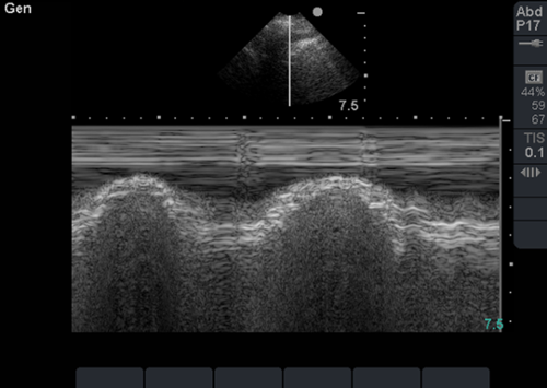
Chest radiograph is pending. In the interim, bedside lung sonography is performed, and the following finding is present on examination of the right lung.
Question 1: The image is consistent with the presence of which of the following:
A. Pleural effusion
B. Pneumothorax
C. Pneumonia
D. Pulmonary embolism
Question 2: The image can be best described as:
A. Bat sign
B. Sinusoid sign
C. Curtain sign
D. Seashore sign
Question 1. Pleural effusion
Question 2. Sinusoid sign.
The sinusoid sign is a dynamic sign seen on M-mode ultrasound when the visceral pleura moves with inspiration decreasing the interpleural distance as the parietal pleura is fixed. The lung moves towards the chest wall with inspiration, the pattern seen on M-mode is a sinusoid. This sign is quiet specific for the presence of a pleural effusion, and can help differentiate small pleural effusions from pleural thickening, which lack the sinusoid sign. It also indicates that the fluid is of low viscosity, as in very viscous or septate effusions the sinusoid sign is absent.
References:
- Lichtenstein DA. General ultrasound in the critically ill. New York: Springer-Verlag 2002.
- Lichtenstein D, Hulot JS, Rabiller A, Tostivint I, Meziere G. Feasibility and safety of ultrasound-aided thoracentesis in mechanically ventilated patients. Intensive Care Med 1999; 25:955-8.
05/2015


