Cough After Visiting the Family Farm
Authors:Thomas Isaacs1,2, Jeffrey Starke1,3, Daniel Hsu1,2
1 Department of Pediatrics, Baylor College of Medicine, Houston, Texas.
2 Division of Pediatric Pulmonology, Baylor College of Medicine and Texas Children’s Hospital, Houston, Texas.
3 Division of Pediatric Infectious Disease, Baylor College of Medicine and Texas Children’s Hospital, Houston, Texas.
Case
An 8-year-old, previously healthy female presents to an emergency room in east Texas with 9 days of dry cough, malaise and fever. Multiple other family members have been ill as well, with symptoms including hemoptysis in the patient’s grandfather. Pertinent exposures include the family chicken coop and a recently abandoned farm (with copious old bird droppings and feathers present) purchased weeks prior to presentation. All sick family members have visited this farm. There are no other pertinent exposures, tuberculosis risk factors, or recent travel. Exam findings include comfortable tachypnea without hypoxemia and normal breath sounds on auscultation. An infectious work-up was performed and chest computed tomography obtained (pictured below).
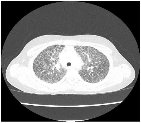 Figure 1: Chest computed tomography (CT) scan
Question
Which additional test result is most likely for this patient?
- Positive interferon-gamma release assay (IGRA)
- Bronchoalveolar lavage (BAL) culture growth of Paracoccidioides
- High serum angiotensin converting enzyme (ACE) level
- Positive histoplasma antigen of urine or serum by enzyme immunoassay (EIA)
D. Positive Histoplasma antigen of urine or serum by enzyme immunoassay (EIA)
Discussion
This patient has disseminated histoplasmosis with severe pulmonary disease, likely secondary to contact with contaminated bird droppings.
Histoplasma capsulatum is a dimorphic fungus which can cause infection through inhalation of microconidia1. Common exposures include bird droppings, chicken coops and buildings undergoing renovation1. Endemic areas include the Ohio and Mississippi River valleys, however more southern areas of the United States, such as Texas and Florida, also have had reported outbreaks1. East Texas is not a coccidioidomycosis or blastomycosis endemic area.
Histoplasmosis can present with a wide range of symptom severity2. Most infections are asymptomatic. When symptoms are present, limited pulmonary manifestations such as cough or dyspnea are most common2. More severe disease may manifest with fever, fatigue, and hypoxemia. Chest radiography may demonstrate lymphadenopathy, infiltrates, or nodules. Occasionally, chronic pulmonary disease will develop which may lead to hemoptysis or cavitary lesions2. A diffuse pattern on imaging is seen with miliary or disseminated Histoplasmosis and is most common with immunocompromised individuals.
Histoplasma antigen testing by EIA can be applied to urine or serum3. Sensitivity and specificity are highest in severe or disseminated disease, and detection can be further improved through a combination of serum and urine testing3,4. Cross-reactivity with other fungal antigens is a limitation of this test. Serology by EIA, immunodiffusion or compliment fixation are alternative non-invasive tests3. Organism isolation from clinical specimens (such as biopsy or BAL) remains the gold standard, however growth may take up to 8 weeks and sensitivity is often low3. The presence of intracellular yeast cells consistent with Histoplasma capsulatum on histopathology can also support the diagnosis3.
Treatment depends on severity. Mild disease that does not last more than four weeks does not require treatment4. For more persistent disease, a six-to-twelve-week course of itraconazole is indicated4. For moderate to severe disease, a one-week course of amphotericin B followed by maintenance therapy with itraconazole for twelve weeks is recommended4. Chronic pulmonary manifestations such as cavitary lesions or mediastinal granulomas may require one to two years of therapy4.
The patient has no relevant exposures to indicate infection with Mycobacterium tuberculosis, making IGRA positivity less likely (answer choice A is incorrect), though with such imaging findings tuberculosis should remain on the differential diagnosis. Paracoccidioides is endemic to Central and South America and initial infection is characterized by systemic symptoms and the absence of pulmonary symptoms, making the finding of Paracoccidioides growth on BAL culture unlikely4 (answer choice B is incorrect). Sarcoidosis is uncommon under the age of 20, and younger children often present with joint pain, uveitis, rash, and a lack of pulmonary symptoms5 (answer choice C is incorrect). Also, the presence of multiple sick contacts highly suggests an infectious etiology. This makes high serum ACE and the diagnosis of sarcoidosis unlikely in this case; however, in adolescents and adults, pulmonary histoplasmosis and sarcoidosis may present similarly5.
References
-
Benedict K, Mody RK. Epidemiology of histoplasmosis outbreaks, United States, 1938–2013. Emerging Infectious Diseases 2016;22(3).
-
Evrard S, Caprasse P, Gavage P, Vasbien M, Radermacher J, Hayette M-P, et al. Disseminated histoplasmosis: Case report and review of the literature. Acta Clinica Belgica 2017;1–8.
-
Azar MM, Hage CA. Laboratory diagnostics for histoplasmosis. Journal of Clinical Microbiology. 2017;55(6):1612–20.
-
Kimberlin DW, Barnett ED, Lynfield R, Sawyer MH, Committee on Infectious Diseases, American Academy of Pediatrics. Red Book: 2021–2024 Report of the Committee on Infectious Diseases.
-
Shetty, Avinash K., and Abraham Gedalia. "Childhood sarcoidosis: a rare but fascinating disorder." Pediatric Rheumatology1 (2008): 1-10.


