A Woman Visiting from China Presents with Shortness of Breath and Fever
Daniel A. Sweeney, MD
Associate Clinical Professor
Division of Pulmonary, Critical Care and Sleep Medicine
University of California, San Diego
La Jolla, CA
History of present illness: An 81 yo woman, visiting from China, presents with two-day history of shortness of breath, fever and chills. She was initially admitted to the general ward but transferred to the ICU for management of hypotension.
Past Medical History: hypertension
Vital signs: BP 90/40 mmHg; pulse 110/min; respiratory rate 22/min; pulse oximetry 94% on 100% NRB mask; temperature 102.1°F
Physical Examination: Tachycardic, Crackles over lower chest bilaterally; abdomen is soft on deep palpation with no rebound or guarding
Initial labs: WBC- 10 x 103/µL; hemoglobin 10.4 g/dl; Creatinine- 1.6 mg/dl; lactate 5.6 mmol/L; urinalysis is unremarkable
Imaging: Portable CXR showed mild cephalization of the vessels and blunting of costophrenic angles bilaterally.
Point-of-care sonography was performed in order to assess volume status and the following representative image was obtained

Question: What is the most likely cause of hypotension in this patient?
- Cardiac failure
- Sepsis
- Dehydration
Answer: B. Sepsis secondary to a liver abscess
This ultrasound movie is a subcostal view of the heart which incidentally reveals a liver abnormality suspicious for an abscess. A subsequent CT was performed which identified a large fluid collection (9x11x11cm) at the junction of the left and right lobes of the liver.
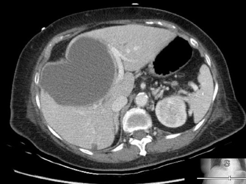
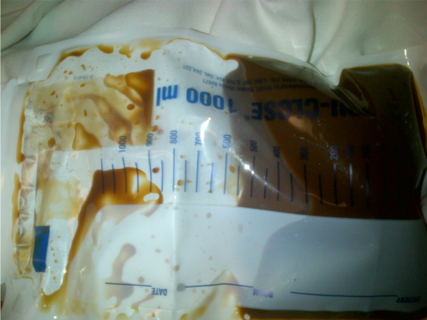
CT-guided placement of an intrahepatic drain was performed and approximately 400ml of brown fluid was immediately aspirated from the fluid collection.
Hepatic abscesses can have a variety of appearances. Abscesses may begin as faint hypoechoic masses. As the abscess evolves it develops a fluid filled center. This fluid can become further complicated by both debris and septations. Abscesses should be differentiated from cysts. Cysts are typically round anechoic structures which demonstrate posterior wall enhancement. Although individual cysts can rupture or become infected, most are benign. In the extreme case of polycystic liver disease—characterized by multiple cysts of varying size—frequent infection or hemorrhage can be problematic. (See Below)
REFERENCE:
- Wang, H.-P., & Chen, S.-C. (2007). Upper abdominal ultrasound in the critically ill. Critical Care Medicine, 35(Suppl), S208–S215


