A Helpful Study
Sarah J Beesley, MD
Pulmonary and Critical Care Medicine Fellow
University of Utah School of Medicine
Case:
A 68 year-old gentleman presented with shortness of breath for 5 days. Review of symptoms was positive for non-productive cough, vomiting and malaise. He denied fevers, chills or chest pain. He had recently been diagnosed with a deep vein thrombosis and started on rivaroxaban. He was admitted to the intensive care unit and a central venous catheter was placed in the right internal jugular vein under ultrasound guidance.
Physical Exam:
T 36.3°C, BP 81/66, HR 119, RR 24, SpO2 92% on 5L nasal cannula. Awake, alert. Heart sounds distant. Respiratory accessory muscle use present.
Relevant Hospital data:
WBC 21.2 k/µL, Creatinine 3.80 mg/dl, Lactate 7.0 mmol/L, INR 9.8, BNP 241 pg/mL, Troponin 0.12 ng/mL. CXR showed right midlung mass.
A study protocol utilizing bedside transthoracic echocardiography (TTE) to evaluate placement of the guidewire in the superior vena cava was performed.
Image 1:
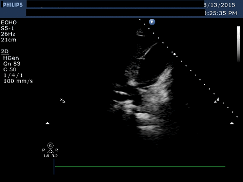
Image 2:
Question:
What is the most likely diagnosis?
Cardiac Tamponade.
On a limited bedside TTE, the patient was found to have a large, generalized pericardial effusion. Diastolic right ventricular and atrial collapse was also present, concerning for cardiac tamponade. A pericardial drain was placed.
Image 1 shows a subcostal window of the inferior vena cava entering into the right atrium. The guidewire is seen within the vena cava. Interposed between the liver and the right ventricle is a large pericardial effusion.
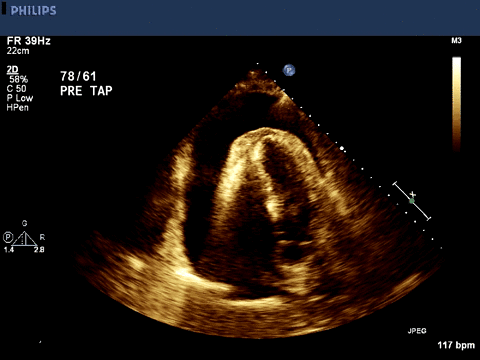
Image 2 shows an apical 4-chamber view, with pericardial effusion and right atrial collapse. The right atrium is the thin-walled chamber on the lower left of the image of the heart. The right ventricular wall is hyperechoic, which obscures some of the distal structures.
Cardiac tamponade is a potentially life-threatening condition due to compression of the heart from a pericardial effusion under pressure that leads to cardiovascular compromise or collapse. Symptoms of cardiac tamponade are generally non-specific and include dyspnea, chest or abdominal pain, and malaise.1 On physical exam, the components of Beck’s triad (hypotension, elevated JVP and muffled heart sounds) are found only in a minority of patients with tamponade.2 Patients are usually hypotensive, tachypneic and tachycardic. Presence of pulsus paradoxus (fall in systolic blood pressure by >10 mmHg during inspiration) increases the likelihood of tamponade.2 While tamponade is often described as being a “clinical diagnosis," confirmatory testing is usually necessary, with echocardiogram being the most common diagnostic test.2
Echocardiogram findings of tamponade include pericardial effusion, usually circumferential, but presence of effusion alone is not indicative of tamponade. Right atrial collapse and right ventricular diastolic collapse is also present. Tamponade can occur regionally as well with localized left heart compression.3 When tamponade is present, left ventricular filling and stroke volume decrease significantly during inspiration (normally there is an initial decrease with inspiration followed by an increase). Additional features include a dilated inferior vena cava without respiratory variation or collapse and abnormal variation in tricuspid and mitral flow velocities.
This patient’s hypotension corrected with drainage of 830 cc of bloody fluid from his pericardium. He remained hemodynamically stable and his pericardial drain was removed 24 hours later. The pericardial fluid had malignant cells present on cytological analysis. He subsequently had a transcutaneous needle biopsy of the right lung mass which was found to be poorly differentiated bronchogenic adenocarcinoma. After consultation with the oncology and palliative care services, the patient was discharged home on hospice.
References:
- Spodick DH. Acute cardiac tamponade. N Engl J Med. Aug 14 2003;349(7):684-690.
- Roy CL, Minor MA, Brookhart MA, Choudhry NK. Does this patient with a pericardial effusion have cardiac tamponade? JAMA. Apr 25 2007;297(16):1810-1818.
- D'Cruz IA, Cohen HC, Prabhu R, Glick G. Diagnosis of cardiac tamponade by echocardiography: changes in mitral valve motion and ventricular dimensions, with special reference to paradoxical pulse. Circulation. Sep 1975;52(3):460-465.


