Sudden Onset of Wheezing at Work
Reviewed By Allergy, Immunology & Inflammation Assembly
Submitted by
Eric J. Adkins, MD
Fellow
Pulmonary, Allergy, Critical Care and Sleep Medicine
The Ohio State University Medical Center
Columbus, Ohio
Matthew Exline, MD
Assistant Professor
Internal Medicine
The Ohio State University Medical Center
Columbus, Ohio
Submit your comments to the author(s).
History
A 33-year-old African American male without significant past medical history, was transferred for evaluation of acute onset of dyspnea (< 48 hours), wheezing and a cough productive of blood-tinged sputum. The patient denied a personal or family history of pulmonary disease. He was previously able to participate in athletic events without symptoms. He denied the use of tobacco, alcohol or drugs. He was employed as an industrial insulation application specialist. Approximately one day prior to presentation, he admitted to an unprotected exposure to a maleic anhydride gas cloud (used as a resin in fiberglass insulation). The patient denied any history of previous exposures. At the time of presentation, the patient did not have a fever or chills and did not report recent weight gain or lower extremity swelling. He had no chest pain, but did complain of chest tightness. He denied nausea, vomiting, diarrhea or abdominal pain. The remainder of his review of systems was unremarkable.
Physical Exam
The patient's weight was 229 pounds. Blood pressure 138/86 mm Hg, pulse 92 beats/minute and oxygen saturation 95% on room air. His initial respiratory rate was 28/min in the Emergency Department and had improved to 22/min at the time of examination. In general, he appeared stable without the use of accessory muscles for respiration. He was able to give us his history without significant dyspnea. Head and neck exam was within normal limits and without evidence of stridor. His lungs revealed fair air movement without wheezing or rhonchi. His cardiac exam was regular rhythm without an S3 gallop or evidence of peripheral edema.
Lab
- Serum chemistry panel and liver function tests were normal.
- Autoimmune serologies including: erythrocyte sedimentation rate (ESR), antinuclear antibodies (ANA) and anti-neutrophil cytoplasmic antibody (ANCA) were negative.
- Complete blood count (CBC) was unremarkable with a normal cellular differential.
- Urinalysis was normal.
Figures
See chest radiograph in Figure 1.
A CT scan of the chest was unremarkable.
Transthoracic echocardiogram during the acute presentation showed mild left ventricular dilation with no evidence of pericardial effusion and an ejection fraction of 25%. A follow-up echocardiogram at 3 months showed resolution of previous abnormalities and a normal ejection fraction.
Pulmonary function testing (PFT) obtained 48 hours after his acute presentation showed a forced vital capacity (FVC) of 3.59 L (88%), a forced expiratory volume in 1 second (FEV1) of 3.10 L (91%), and an FEV1 / FVC of 86.4%. Lung volumes were normal. The diffusing capacity for carbon monoxide was 59%. On 6-minute walk testing, he ambulated 1658 feet with an oxygen saturation of 98% at rest and 94% during exercise.
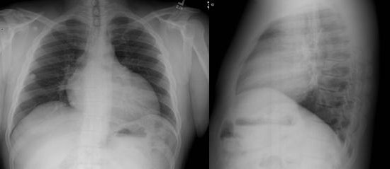
Figure 1. PA and lateral chest radiographs from the patient. The images show mild elevation of right hemi-diaphragm, normal lung fields and cardiomegaly.
RADS is a term used to describe bronchial hyper-reactivity after exposure to an inhaled irritant. It may also be known as irritant-induced asthma as described in a spectrum of work related asthma entities (1). RADS was first described in 1981 and later in more detail in 1985 after a review of ten subjects who all presented with an acute onset of asthma-like symptoms after exposure to an inhaled chemical irritant (2, 3). It is primarily a clinical diagnosis that depends significantly upon a detailed and accurate history (4). One key to the diagnosis is the temporal relationship between an appropriate exposure and the development of symptoms. Patients often present after a known massive exposure that often prompts them to seek immediate medical attention. The initial injury in RADS is the inhaled irritant causing direct damage to the bronchial mucosa with subsequent epithelial cell necrosis and proliferation of the basement membrane. Bronchial biopsies later in the disease course reveal desquamation of the bronchial epithelium with a chronic lymphocytic infiltrate without evidence of mucus gland hypertrophy or smooth muscle hyperplasia (4). Table 1 describes the criteria needed to make a diagnosis of RADS.
- Documented absence of preceding respiratory complaints
|
- The onset of symptoms occurred after a single specific exposure incident or accident
|
- The exposure was to a gas, smoke, fume or vapor which was present in very high concentration and had irritant qualities to its nature
|
- The onset of symptoms occurred within 24 hours after the exposures and persisted for at least 3 months
|
- Symptoms simulate asthma with cough, wheezing and dyspnea
|
- PFT may show airflow obstruction.
|
- Methacholine challenge testing is positive
|
- Other pulmonary diagnoses have been excluded
|
Table 1. Diagnostic Criteria for RADS (2, 3)
The other diagnostic possibilities are unlikely given this presentation. The acute onset with known exposure without any previous pulmonary symptoms makes a diagnosis of asthma less likely. The patient’s lack of hemoptysis or anemia and a clear chest radiograph makes a diagnosis of alveolar hemorrhage unlikely. A diagnosis of VCD would not explain the abnormal PFTs nor the acute presentation. Hypersensitivity pneumonitis would generally be associated with low-level chronic exposure rather than an acute massive exposure and would be associated with an abnormal chest radiograph.
The presence of bronchial hyper-reactivity is an essential element to making a diagnosis of RADS. Methacholine is similar to acetylcholine, but its effects last longer allowing for changes in airflow to be detected. When interpreting the test, it is the provocative concentration that decreases the FEV1 by 20% (PC20) that is used to quantify the degree of bronchial hyper-responsiveness. Cutoff values for determining the presence of hyper-reactivity are shown in Table 2.
Bronchoscopy with bronchoalveolar lavage would rarely be indicated in the setting of normal lung fields on the chest radiograph. The use of inhaled corticosteroid can be useful in the treatment of RADS. However, methacholine challenge should be done to confirm the diagnosis first. The patient does not have VCD, so laryngoscopic evaluation would not be appropriate at this time. Occupational medicine may be valuable in this patient at some time, but would not be helpful in establishing the diagnosis of RADS at this time.
PC20 (mg/ml) |
Interpretation
|
|
>16
|
Normal bronchial responsiveness
|
|
4.0-16
|
Borderline bronchial hyper-reactivity (BHR)
|
|
1.0-4.0
|
Mild BHR (positive test)
|
|
<1.0
|
Moderate to severe BHR
|
Table 2. Methacholine bronchial responsiveness categories (5)
There is no data from randomized controlled trials to suggest the best management strategies for treatment of RADS. Most of the data supporting treatment of RADS comes from case reports. One case series of four patients diagnosed with RADS related to occupational exposures described improvement with treatment of inhaled steroids and bronchodilators (6). Patients with an established diagnosis of RADS should be treated with inhaled corticosteroids. Persistent symptoms of bronchospasm should receive treatment with short-acting beta-agonists as needed.
In patients who may have been exposed to the offending agent from occupational exposure, all efforts must be made to prevent re-exposure. Patients should be urged to seek out alternative employment or different duties with the same employer to minimize any risk of recurrence. If they remain employed in the same setting of the original exposure, all respiratory protective methods recommended at the worksite should be followed at all times.
While any inhaled gas, smoke, fume or vapor can be an inciting cause of RADS, chlorine has been found to be the most common offending agent. In a recent systematic review of published cases in which the criteria for RADS were present, chlorine exposure was found to the most prevalent (7). Other agents that were noted to be causative were toluene di-isocyanate and nitrogen oxides.
In the same review of RADS, the workplace was the most common setting for exposure to an irritant agent and onset of symptoms. It is important to realize that RADS can occur in other environments outside of the work setting. A detailed history that includes any possible exposures a patient may have had at home or in other environments is essential towards making a diagnosis of RADS when it is considered.
Follow Up:
Our patient’s methacholine challenge revealed mild bronchial hyper-reactivity with a provocation concentration with 20% reduction in FEV1 (PC20) of 3.13 mg/ml. He was treated with daily inhaled corticosteroids and a beta agonist inhaler as needed. With therapy, his dyspnea with intermittent cough and wheezing slowly resolved over a period of 6 months. Our patient chose to remain with his employer and attempted to return to work. However, mere entrance to the work area without any respiratory protection re-exposed him to the inciting agent resulting in prompt recurrence of his signs and symptoms. He was unable to return to his previous occupation.
References
- Tarlo SM, Balmes J, Balkissoon R, Beach J, Beckett W, Bernstein D, Blanc PD, Brooks SM, Cowl CT, Daroowalla F, Harber P, Lemiere C, Liss GM, Pacheco KA, Redlich CA, Rowe B, Heitzer J. Diagnosis and management of work-related asthma: American College Of Chest Physicians Consensus Statement. Chest 2008;134:1S-41S.
- Brooks SM, Lockey J. Reactive airways disease syndrome (RADS): a newly defined occupational disease [abstract]. Am Rev Respir Dis 1981; 123 (suppl):A133.
- Brooks SM, Weiss, MA, Bernstein, IL. Reactive airways dysfunction syndrome (RADS): Persistent asthma syndrome after high level irritant exposures. Chest 1985; 88(3):376-384.
- Alberts WM, do Pico GA. Reactive airways dysfunction syndrome. Chest 1996;109:1618-1626.
- American Thoracic Society. Guidelines for methacholine and exercise challenge testing-1999. Am J Respir Crit Care Med 2000; 161:309-329.
- Matrat M, Laurence MF, Iwatsubo Y, Hubert C, Joly N, Legrand-Cattan K, L’Huillier JP, Villemain C, Pairon JC. Reactive airways dysfunction syndrome caused by bromochlorodifluoromethane from fire extinguishers. Occup Environ Med 2004 Aug;61(8):712-714.
- Shakeri MS, Dick FD, Ayres JG. Which agents cause reactive airways dysfunction syndrome (RADS)? A systematic review. Occupational Medicine 2008;58:205–211.


